SEM and EDS Fracture Analysis Limitations: Association Is Not Cause
Practical reading for undercarriage components, operating conditions and parts decisions.
On this page
Scanning electron microscopy (SEM) and energy-dispersive X-ray spectroscopy (EDS) can show what a fracture surface looks like and what elements appear at a chosen spot. They do not, on their own, identify why a part failed. An oxygen peak, a sulfur signal or an inclusion near an origin is an observation tied to one location. It becomes part of a cause conclusion only when it is linked to a fracture map, the part's handling history and an interpretation basis, and when other evidence supports it. Frame the investigation so that every image and spectrum can be traced and every cause statement stays conditional until corroborated.

What each technique contributes
Element, a testing laboratory, describes SEM as a tool for determining the origin, mode and direction of propagation of cracks or fractures. It says SEM is used together with macro photography and stereomicroscope documentation to give a complete representation of the features examined. Images come from secondary electrons, which give finer surface resolution, and backscattered electrons, which give topographic detail and information related to atomic weight. The same article notes that SEM images lack color, that samples must be electrically conductive or treated to manage that, and that SEM magnification runs from roughly 5X to over 50,000X.
EDS is the chemistry layer. Element says it provides the relative chemical composition of the surfaces and features examined and can be combined with images into elemental maps that show how elements are distributed. It describes uses such as identifying corrosion products, residues, inclusions and wear debris. That is a laboratory's own description of its method, not an independent standard.
The key word is relative. EDS reports what is present at the analyzed spot on the analyzed surface. It does not report the bulk alloy of the part, and it cannot by itself say whether a surface element was in the steel, came from the environment or was added during handling.
Standards-based and standardless results are different
EDS results can be calculated with reference standards or without them. NIST lists a publication on standardless quantitative electron-excited X-ray microanalysis by energy-dispersive spectrometry, which shows that standardless quantification is a distinct basis with its own accuracy question. The retrieved NIST page gave only the listing, so this article draws no numerical error from it. The practical step is to ask the laboratory which basis it used, and to treat a standardless composition as an estimate for the examined feature, not as proof of a material grade.
Build a fracture feature ledger
Start from the preserved part and its fracture map, then record each analyzed feature on its own line. Evidence recovery and packing belong to preserving a failed metal part for laboratory examination. The ledger begins after that step. It is a recommended review aid, not a form required by any standard.
Keep observation, interpretation and cause apart
A single observation can fit several explanations. An oxygen-rich region on a fracture face could reflect an oxide that formed in service, an oxide that formed afterward during storage or a contaminant. A sulfur-bearing feature near an origin could be an inclusion that initiated the crack or one that happened to be exposed. The ledger should hold these as hypotheses with the evidence for and against each.
Corroboration comes from outside the microscope. Service history, loading conditions, the manufacturing record, hardness and microstructure examination and chemistry of the bulk material each test a hypothesis from a different direction. A cause statement is ready when independent lines of evidence agree and the alternatives have been addressed. Where they do not, the correct entry is "undetermined," with the missing evidence named.
Systematic cleanliness classification is a different task. For how inclusion ratings are read across a steel, see what steel cleanliness reports can prove. One failed fracture feature does not substitute for it.
Write the request and read the report
Ask the laboratory for the investigation in the ledger's terms: the mapped regions to be examined, the imaging and EDS modes, the quantification basis, the original images and spectra, and an explicit statement of which conclusions the evidence supports and which it does not. When the report arrives, check each cause statement against the ledger. Any statement that rests on one spectrum, or that ignores a competing explanation, should go back as unresolved.
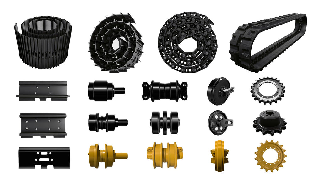
For a part supplied by KTSU, provide the part reference, the failure description and the preserved-evidence record when asking what documentation is available. The public excavator undercarriage parts collection identifies the product family. It does not establish that KTSU performs SEM or EDS work or reaches failure conclusions.
The review is complete when every feature has a place in the register, each observation is separated from its interpretation, and the corroboration needed before any cause is stated is named.
More Technical Guides guides
Find the next article for your component, application or buying decision.
Back to Technical Guides